
The NDRL is committed to producing research with translational applications to parents, educators, and clinicians who work with children. By partnering with local organizations, schools, and clinics in the Hongkong, we hope to expand the impact of our research beyond PolyU to improve the lives of children with developmental and learning disabilities. This page is dedicated to sharing resources with parents and educators looking to learn more about Developmental Dyslexia (DD), Developmental Language Disorder (DLD), and Autism Spectrum Disorder (ASD).
Have a suggestion to add to our page? We are always happy to receive feedback, and would gladly add further resources or recommended readings to our page. Please contact with suggestions for additions to our resources page!
1. Lab Tools


Functional magnetic resonance imaging (fMRI)
Functional magnetic resonance imaging or functional MRI (fMRI) measures brain activity by detecting changes associated with blood flow.[1][2] This technique relies on the fact that cerebral blood flow and neuronal activation are coupled. When an area of the brain is in use, blood flow to that region also increases.[3]
The primary form of fMRI uses the blood-oxygen-level dependent (BOLD) contrast,[4] discovered by Seiji Ogawa in 1990. This is a type of specialized brain and body scan used to map neural activity in the brain or spinal cord of humans or other animals by imaging the change in blood flow (hemodynamic response) related to energy use by brain cells.[4] Since the early 1990s, fMRI has come to dominate brain mapping research because it does not involve the use of injections, surgery, the ingestion of substances, or exposure to ionizing radiation.[5] This measure is frequently corrupted by noise from various sources; hence, statistical procedures are used to extract the underlying signal. The resulting brain activation can be graphically represented by color-coding the strength of activation across the brain or the specific region studied. The technique can localize activity to within millimeters but, using standard techniques, no better than within a window of a few seconds.[6]

Mock MRI
An MRI mock scanner simulates the real MRI experience, including the noises the camera makes throughout the scan. The mock scanner is a useful tool to see how your child will handle the MRI and assess their ability to complete an MRI without sedation. The purpose of the Mock MRI is to enable both children and adults who require an MRI scan to become familiar with the scanner environment, and to practice any tasks expected of them (like keeping still or holding their breath).
A certified child life specialist will set up the mock scan and walk your child through the process. If your child is allowed to watch or listen to a video or music during their scan, the mock scanner is equipped to simulate these options as well.

EEG (electroencephalogram)
An electroencephalogram (EEG) is a test that measures electrical activity in the brain. This test also is called an EEG. The test uses small, metal discs called electrodes that attach to the scalp. Brain cells communicate via electrical impulses, and this activity shows up as wavy lines on an EEG recording. Brain cells are active all the time, even during sleep.
An EEG is a valuable tool that can detect changes in brain activity, aiding in the diagnosis of various brain conditions. It is particularly useful for identifying epilepsy and other seizure disorders. Beyond this, EEG can assist in diagnosing or treating brain tumors, brain damage from head injuries, encephalopathy (a brain disease with diverse causes), brain inflammation like herpes encephalitis, strokes, and sleep disorders. It can even help in diagnosing Creutzfeldt-Jakob disease. Additionally, EEG might be employed to confirm brain death in comatose individuals and, through continuous monitoring, can help determine the appropriate level of anesthesia for those in a medically induced coma.


fNIRS (Functional near-infrared spectroscopy)
Functional near-infrared spectroscopy (fNIRS) is an optical brain monitoring technique which uses near-infrared spectroscopy for the purpose of functional neuroimaging.[1] Using fNIRS, brain activity is measured by using near-infrared light to estimate cortical hemodynamic activity which occur in response to neural activity. Alongside EEG, fNIRS is one of the most common non-invasive neuroimaging techniques which can be used in portable contexts. The signal is often compared with the BOLD signal measured by fMRI and is capable of measuring changes both in oxy- and deoxyhemoglobin concentration,[2] but can only measure from regions near the cortical surface. fNIRS may also be referred to as Optical Topography (OT) and is sometimes referred to simply as NIRS.
fNIRS estimates the concentration of hemoglobin from changes in absorption of near infrared light. As light moves or propagates through the head, it is alternately scattered or absorbed by the tissue through which it travels. Because hemoglobin is a significant absorber of near-infrared light, changes in absorbed light can be used to reliably measure changes in hemoglobin concentration. Different fNIRS techniques can also use the way in which light propagates to estimate blood volume and oxygenation. The technique is safe, non-invasive, and can be used with other imaging modalities.
2. Highlights
Leading the way in AI and humanities research
As AI brings new possibilities to all corners of society, The Hong Kong Polytechnic University is at the forefront of digital advances in disciplines such as the humanities, languages, health, neuroscience and history.
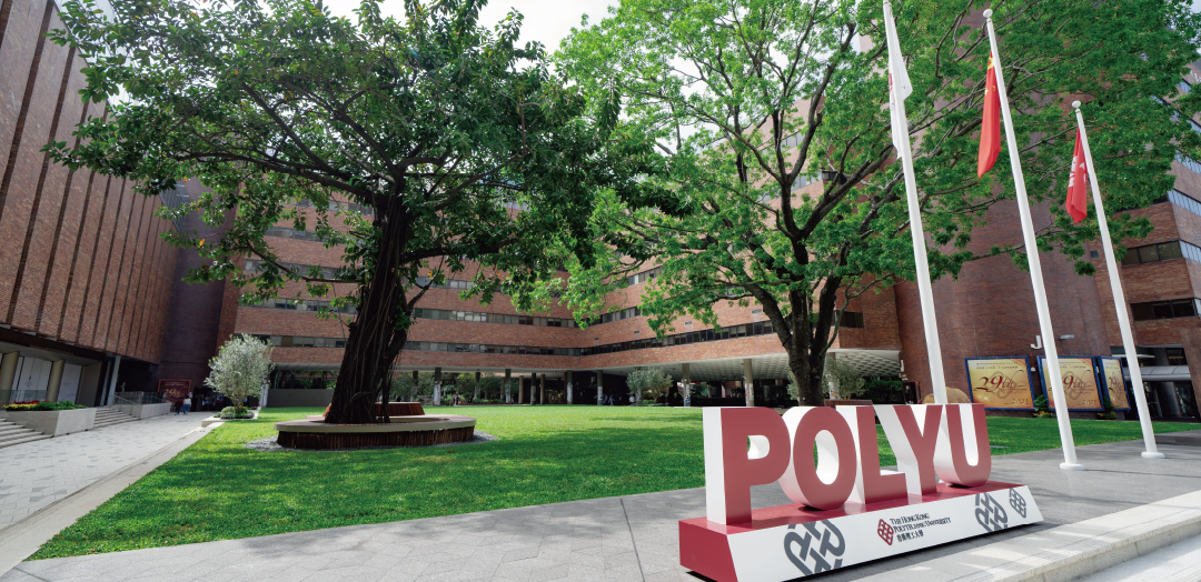
The Hong Kong Polytechnic University (PolyU) is an innovative, world-class university with more than 85 years of rich heritage. It aims to nurture socially responsible professionals and leaders and pursue world-leading research and innovation for societal benefits, in line with the university’s motto: “To learn and to apply, for the benefit of mankind.” Through cutting-edge research that brings together the humanities and modern technology, PolyU’s impact stretches beyond Hong Kong.
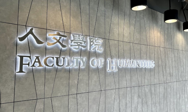
Building on forward-looking initiatives at the university, the Division of Artificial Intelligence and Humanities in PolyU’s Faculty of Humanities – set to launch in January 2026 – will serve as a catalyst for the Faculty’s transition towards AI integration, bridging technological expertise with the full spectrum of humanities disciplines.
3. Technical Explanation
This is a animated explanation, explaining an fMRI experience from a child’s perspective, with a demonstration of the relevant process (in animated form).
这个是一个动画片的讲解,从小朋友的角度讲解自己的一次fMRI经历,有相关流程的演示 (动画形式)。
This is a video of a real-life experiment, showing the entire process of a child undergoing fMRI.
这个是真人实验的流程拍摄,展示了一个小朋友做fMRI的全流程。
A comprehensive introduction to MRI equipment, in easy-to-understand animated format.
全面的MRI设备科普,动画的形式通俗易懂。
The entire process is an introduction to fNIRS presented in the form of animated pictures.
全程是动画图片的形式呈现对fNIRS的介绍。
This video introduces fNIRS using animations and images, describing the relationship between fNIRS and MRI (why fNIRS is performed after MRI) and explaining the operating principles and advantages of fNIRS.
动画和图片的形式介绍了fNIRS,叙述了了fNIRS和MRI的关系(为什么做了MRI还要做fNIRS)。讲述了fNIRS的操作原理和优势等。
4. Education Blogs


[Lab for NDRL] What Every Parent Should Know About Developmental Dyslexia
Ralph Liu @ NDRL – Feb 27, 2024
[Child Assessment Service] Developmental Disorders in Children – Developmental Language Disorder (DLD)
Tegan Yeung @ NDRL – Feb 24, 2024

[Child Assessment Service] Developmental Disorders in Children – Dyslexia
Tegan Yeung @ NDRL – Feb 26, 2024

[Child Assessment Service] Developmental Disorders in Children – Autism Spectrum Disorder (ASD)
Tegan Yeung @ NDRL – Feb 23, 2024
4. Useful Links


